

Optical Microscopy
Stereomicroscopy is used for low magnification applications, allowing high-quality 3D observation of materials that are normally visible to the naked eye. Stereoscopic microscopes observe the sample through two different lenses. This allows the image that reaches each eye to be slightly different. The combination of these two images through our eyes produces the three-dimensional effect.
The Fluorescence Microscopy technique allows, thanks to an optical microscope that has a specific architecture, the detection of specific areas of the cell to which the fluorescent tracers that we previously added to the samples have attached.
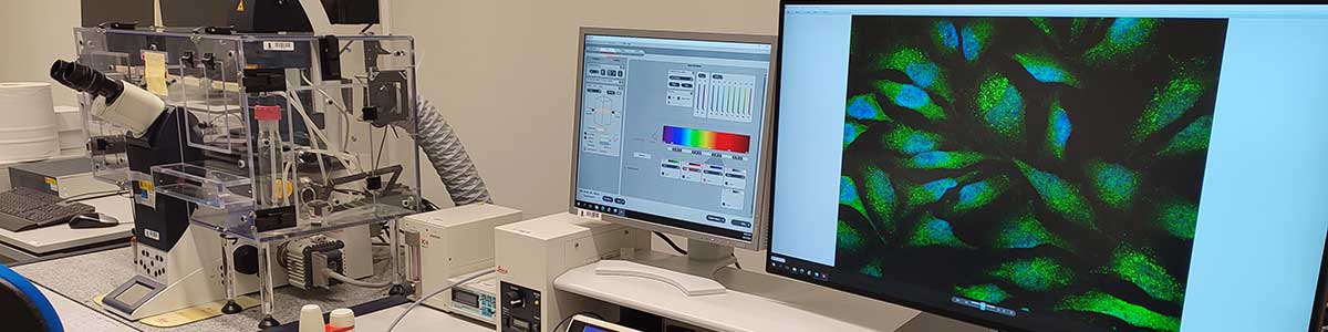
Confocal Microscopy also uses fluorescent tracers but, unlike Fluorescence Microscopy, only the fluorescent emission from the focused area is collected, rejecting the rest and thus achieving an increase in resolution.
This, together with the fact of having an incident laser light, enables this microscope to acquire hundreds of very thin confocal optical sections in different planes of the sample, which can be stored and combined to generate a three-dimensional image in a non-destructive way. The image view can be of the whole or of certain parts.
For its part, the fact of having an incubation chamber allows work on live cells, with the possibility of collecting very fast images, both in Wide Field mode and in confocal mode thanks to the high-frequency scanner.
Instrumental Facilities
Nikon SMZ1500 Zoom Stereoscopic Microscope
- CW10xA/22 eyepieces
- HR Plan Apo 1x WD54 Objective
- Parallel optical system with zoom.
- Zoom range: 0.75x-11.25x
- DeltaPix Infinity X Camera
- Software DeltaPix InSight
- Superior illumination and epifluorescence with GFP-B cube (EX 480/40 DM505 BA 535/50)
Nikon Eclipse E800 Microscope
Objectives:
- 4xPL Fluorine 0.13
- 10x PL APO 0.45
- 20x PL Fluor 0.50
- 40x PL APO 1.0 OIL
- 60x PL APO 1.4 OIL
- 100x PL APO VC 1.4 OIL
Nikon DS-5 Mc CCD camera
- Epifluorescence module:
- B2A. Blue excitation
- G2A. Green Excitation
- V2A. Ultraviolet Excitation
- Dell Workstation
- Polarizer and Analyzer
- LINKAM SCIENTIFIC PE120 Temperature Plate
Leica SP5 Confocal Module on Leica DMI 6000 Inverted Optical Microscope
Objectives:
- 10x PL APO 0.40
- 20x PL APO 0.70
- 20x PL APO 0.70 IMM
- 40x PL APO 0.75 OIL
- 63x PL APO 1.4 OIL
- 63x HCX PL APO 1.20 Water
DIC in all objectives
Fluorescence Filters:
- I3. Blue Excitation
- N2.1. Green Excitation
- UV excitation
- Galvanometric plate, motorized in X and Y
- Incubation plate
- Leica SP5 Multispectral Confocal Module with 3 photomultiplier detection channels, one channel with hybrid detector and a fifth for transmission
Laser equipment:
- Blue, Argon (458nm, 476nm, 488nm, 496nm, 514nm)
- Verde, HeNe (543nm)
- Red, HeNe (633 nm)
- Violet, Blue Diode 405nm)
- Acoustic-optical splitter (AOBS)
- Multidimensional series acquisition: x, y, z, t, o, λ
- Software FRAP, FLIP, FRET, 3D, Dye Finder
- Incubation chamber with CO2 and temperature control
- Water dispenser for lenses
- Wide Field
- High Content Screening image collection system in confocal mode (Matrix software)
- Metamorph image collection and processing software
Applications
- Marking by immunofluorescence of membrane proteins, subcellular organs, cytoskeleton, etc.
- Cellular physiology studies
- Reconstruction of three-dimensional structures
- Study of micro-roughness in polished materials
- Analysis of samples with fluorescence of their own
- Live cell studies
- High Content Screening
Sample requirements
Contact Technical Staff
More information
María Inés Pazos Garrido – Beatriz Rivas Murias – Catalina Sueiro López – Daniel Carlos Cernadas Fraga
+34 986 812 125 +34 986 812 110
inespazos@uvigo.gal – brivas@uvigo.gal – catalina.sueiro@uvigo.gal – daniel.carlos.cernadas.fraga@uvigo.gal