

Single Crystal X-Ray Diffraction
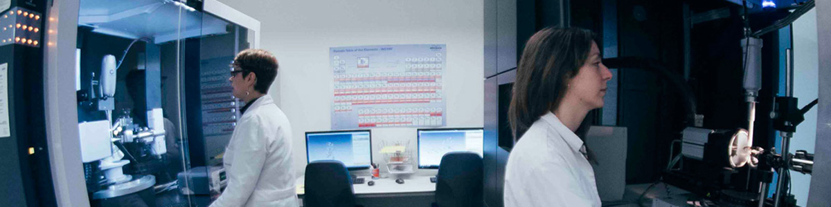
‘Single Crystal X-Ray Diffraction’, commonly known as ‘X-Ray Crystallography’, is an analytical technique in which X-ray methods are used to determine with certainty the actual arrangement of atoms within the crystal under study and, therefore, the three-dimensional ionic or molecular structure as well as the supramolecular organization. The science of ‘X-Ray Crystallography’ originated in 1912 with Laue’s discovery that crystals diffract X-rays. Since then, single crystal X-ray diffraction has become the most effective tool for obtaining the spatial distribution of atoms in the solid state.
Structure determination by X-ray diffraction can be applied to a wide range of systems, from very small molecules or simple salts, to complex minerals, synthetic organic, inorganic and organometallic compounds, natural products and biological macromolecules, such as proteins and even viruses. Precise knowledge of molecular geometry is becoming increasingly important in almost all fields of chemical and biological research as it allows understanding both the physical properties – color, electrical conductivity, magnetic properties -, chemical properties – reactivity, catalytic properties – and/or biological properties – pharmacological properties – of the systems under study.
The Single Crystal X-ray Diffraction Unit is equipped with the necessary equipment to carry out structural studies of ‘small molecules’. ‘Small molecule’ crystallography typically involves crystals with less than 100 atoms in their asymmetric unit.
Instrumental Facilities
Bruker D8 Venture Diffractometer
Equipped with:
- Detector bidimensional CMOS Photon 100
- Kappa type four-circle goniometer
- 50 W Xerador with high-brillo molybdenum microfonte
- Helios™ multilayer Göbel optics (model ELM33)
- CryoStream 800 Low Temperature Unit
Year of acquisition: 2015
Nikon SMZ-745T Stereoscopic Microscope
Equipped with:
- Hybrid LED Lighting Stand
- Oculares C-W 20x
- Nikon P6000 digital camera
Year of acquisition: 2009
Nikon SMZ-745T Stereoscopic Microscope
Equipped with:
- Hybrid LED Lighting Stand
- Oculares C-W 20x
- Nikon P6000 digital camera
Year of acquisition: 2013
Applications
- Measurement of the crystal’s lattice parameters.
- Identification of known substances through reticular parameters.
- Determination of the three-dimensional ionic or molecular structure: Atomic positions, distances and bond angles, torsion angles, etc.
- Evaluation of supramolecular interactions (hydroxene bond, π···π , CH···π , haloxene···haloxene) related to the physical properties of the materials studied.
- Determination of absolute configuration for enantiopure samples.
- Estimation of phase transitions from measurements at variable temperature.
The group of areas that have traditionally shown interest in this technique, such as Mineralogy, Inorganic Chemistry, Organic Chemistry and Solid State Chemistry, are joined by others such as Materials Science, Pharmacology, Natural Products, Biochemistry, etc.
Sample requirements
The samples must consist of single crystals with defined faces and edges, without striations, inclusions or dislocations, the preferable size being approximately 0.15 x 0.15 x 0.15 mm. To avoid the degradation of unstable crystals, it is advisable to send them submerged in mother liquors or protected with mineral oil.
More information
Berta Covelo
+34 986 812 669